
Related Post

How Cryo-EM Captures Protein Dynamics
2025-07-30Proteins aren’t statues – they move, twist, fold, and change shape constantly. Understanding this motion is key to unlocking how life works at the molecular level. And that’s where Cryo-EM enters the picture. This advanced imaging technique is making it possible to capture proteins not as static structures, but in motion – frame by frame – offering a clearer window into their function. At Longlight Technology, we’ve witnessed firsthand how Cryo-EM is reshaping structural biology. As a manufacturer of precision laboratory equipment, we’re proud to support this scientific revolution with tools designed for clarity, speed, and accuracy.
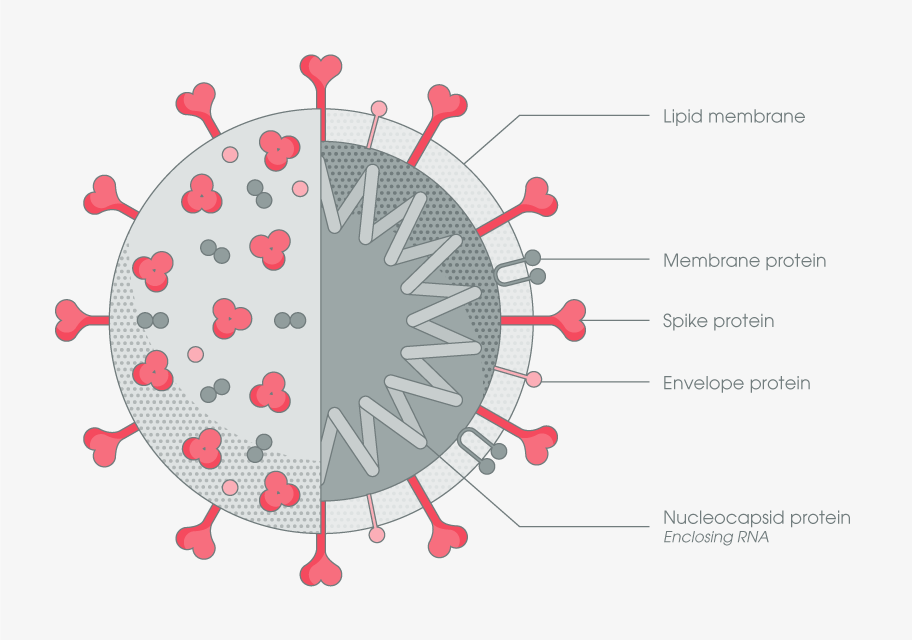
(Conformational dynamics capture of the SARS-CoV-2 spike protein)
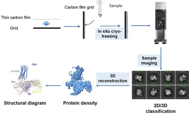
Cryo-Electron Microscopy (Cryo-EM) is an essential structural biology tool widely applied in cutting-edge biomedical research. For example, it was used to capture the conformational dynamics of the SARS-CoV-2 spike protein, which was critical for understanding viral entry mechanisms and directly supported the design of vaccines and neutralizing antibodies during the COVID-19 pandemic (Wrapp et al., Science, 2020). Another major application lies in studying protein misfolding and amyloid fibril formation in neurodegenerative diseases like Alzheimer’s disease, helping researchers elucidate the molecular basis of pathological aggregation (Fitzpatrick et al., Nature, 2017). Cryo-EM enables visualization of large and dynamic protein complexes at near-atomic resolution without the need for crystallization, making it indispensable for studying complex biological processes in their native states.
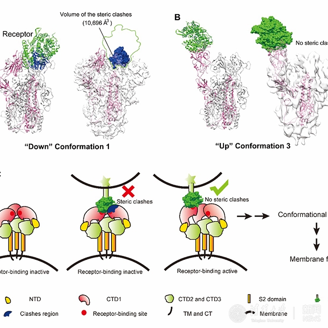
(Tsinghua University research teams led by Wang Xinquan and Xiang Ye published a study revealing a key step in coronavirus entry into host cells.)
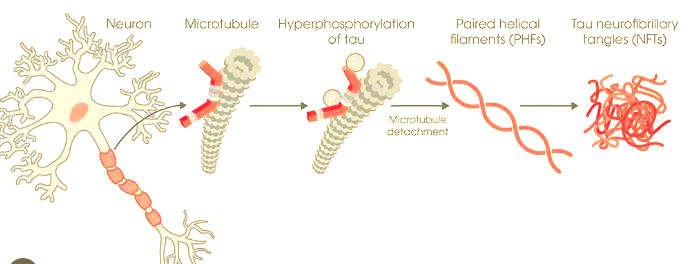
(Alzheimer’s Disease and Prion Diseases: Striking Similarities in Protein Folding and Propagation)
Watching Molecules in High Resolution
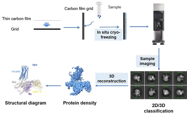
For decades, scientists have sought tools that could reveal how biological molecules behave – not just what they look like. Traditional cryo-electron microscopy (Cryo-EM) brought us closer to this goal by allowing researchers to freeze delicate biomolecules in their native, hydrated state and study them at near-atomic resolution. Unlike crystallography, which requires fixed, uniform structures, Cryo-EM works with complex, flexible proteins – no crystallization needed.
Cryo-EM has revolutionized structural biology by enabling visualization of large, dynamic protein complexes in their native conformation. Rather than offering a single fixed view, advanced image processing techniques applied to Cryo-EM datasets allow researchers to computationally reconstruct multiple structural states from heterogeneous particle populations. This reveals conformational variability that was previously invisible – giving insights into molecular mechanisms, protein-ligand interactions, and allosteric regulation.
This perspective has massive implications:
✅Complex enzymes can be visualized in different conformational states, helping clarify catalytic mechanisms.
✅Transient intermediate structures can be inferred from particle heterogeneity, offering a deeper understanding of protein function.
✅Ligand binding, conformational switches, and multi-domain arrangements can be studied under native-like conditions, such as physiological pH and temperature.
By allowing scientists to visualize molecular architecture with exceptional clarity, Cryo-EM fills a long-standing gap in structural biology. Instead of relying solely on crystallized snapshots or indirect models, researchers can now study biomolecular machines in action, even if only inferred from equilibrium mixtures. That insight isn’t just academically satisfying – it has practical value, too. From drug development to synthetic biology, understanding macromolecular structure under near-native conditions opens doors to smarter, faster innovation.
Why This Technology Is a Big Deal
In the world of protein research, structural flexibility is often key to function. Many proteins shift between conformations as they bind to molecules, process substrates, or transmit signals. Capturing these different states – especially for membrane proteins, viral fusion proteins, or multi-subunit complexes – has been challenging for traditional methods.
Cryo-EM changes that. It gives researchers the ability to freeze biomolecules in their functional states – without distorting their natural form. This is a massive step forward for structural biology, especially when studying proteins that are unstable, disordered, or exist in multiple configurations.
What makes this method truly game-changing is its practicality:
- Work with nanoliter-scale sample volumes, saving precious biological material
- Skip the long, difficult crystallization process entirely
- Preserve the native hydration state, keeping molecules biologically relevant
- Resolve heterogeneous conformations from a single dataset
These advantages open up new possibilities for studying everything from viral entry mechanisms to motor proteins and signaling enzymes. In drug discovery, where understanding the exact binding conformation of a molecule can determine therapeutic success, Cryo-EM offers essential structural insights that static methods often miss.
At Longlight Technology, we’ve built our Cryo-EM systems with these needs in mind. Our platforms support ultra-fast vitrification, high-contrast imaging, and precision control to capture every structural detail. Whether you’re exploring enzyme structure or protein misfolding in disease, our tools help make the invisible visible – with clarity, accuracy, and confidence.
Behind the Tools at Longlight Technology
As a company, we’re not just focused on machines – we’re focused on the future of science. At Longlight Technology, we design and manufacture high-end laboratory instruments, genomic consumables, and structural biology solutions tailored to real-world research needs.
Our commitment goes beyond providing hardware. When it comes to Cryo-EM, we offer:
- Complete Workflow Support – From vitrification to data analysis, we’re with you at every step
- Flexible Imaging Solutions – Whether you’re doing single-particle analysis or tomography
- Expert Assistance – Our experienced team helps optimize your experimental design and output
Beyond Cryo-EM, our catalog includes focused ultrasonication systems, nucleic acid extraction kits, ChIP-seq consumables, and various NGS preparation tools. Every product is built with reliability and precision in mind, supporting fields from basic biology to clinical diagnostics.
Closing Words
We’re entering a new era in molecular visualization. With Cryo-EM, researchers no longer need to rely solely on indirect or static snapshots – they can analyze structural heterogeneity and conformational states with near-atomic resolution. That opens doors to new discoveries in everything from enzyme mechanisms to vaccine design. We believe this technology will play a central role in understanding how biology truly works – in space, structure, and complexity. As manufacturers, we take pride in knowing that our instruments help push those boundaries forward. We’ve already seen Cryo-EM help labs uncover protein architectures that were previously inaccessible – and that’s just the beginning. If you’re ready to explore the frontiers of molecular structure, Longlight Technology is here to equip your lab with the tools to do it. Let’s build discoveries that move the world.