Cryo‐electron Microscopy Service
Cryogenic electron microscopy (cryo-EM) is a cutting-edge technique in structural biology that allows scientists to visualize the precise details of biological macromolecules. Cryo-EM is based on transmission electron microscopy at near atomic resolution. Increasing market demand in numerous biomedical applications, e.g. drug development, antibodies, vaccines and so on.
Advantages of cryo-EM over X-ray crystallography
Despite the X-ray diffraction still can reach slightly greater resolution, cryo-EM exhibits significant advantages:
- Minimal amount of sample is needed.
- No need for crystallization.
- No need for synchrotron source of X-ray.
- Preservation of near-native state.
- Evaluation of the flexibility.
- Suitable for particles that are difficult to crystallize such as membrane proteins or supramolecular assemblies (e.g. virus).
- The heterogeneity of conformations can be resolved in a single specimen.
- Low level of purity accepted.
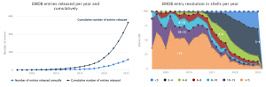
Exponential growth of the number of entries in EMDB:
Structural Solutions
- Sample Suitability Assessment – Negative Stain
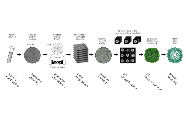
Prior to committing important microscope resources in cryo-EM, negative stain examination is generally performed at room temperature and low voltage. This step allows the assessment of sample suitability, including homogeneity, aggregation, particle morphology, size, and distribution, using a high-electron-density staining agent.
Negative staining serves as a critical quality control step to evaluate whether a sample is appropriate for downstream cryo-EM data acquisition.
- High-Resolution Structure Determination – Single Particle Analysis (SPA)
Cryo-EM data acquisition typically generates approximately 10⁴ to 10⁶ individual two-dimensional projections of particles captured at various orientations. These projections are computationally aligned and classified through an image processing workflow to reconstruct a three-dimensional density map at sub-nanometric or near-atomic resolution.
An atomic model is subsequently built, refined, and validated within the reconstructed density map, providing structural insights into the functional mechanisms of the biological macromolecule of interest.
- In Situ Structural Analysis – Cryo-Electron Tomography (Cryo-ET)
Cryo-electron tomography enables three-dimensional visualization of biological structures in their native cellular or viral context. This approach is particularly suitable for studying large macromolecular assemblies, membrane-associated complexes, and viral infection mechanisms, offering spatial and organizational information inaccessible by single-particle methods.
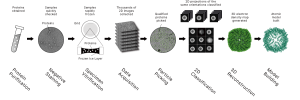
Service Workflow
Project consultation → NDA signing → service agreement confirmation → sample receiving → quality inspection → negative stain screening → cryo-EM data collection → data processing → report delivery.
Data & Result Deliverables
Full data availability is provided throughout the project lifecycle. Delivered materials include raw cryo-EM movies with gain reference files, intermediate data files at various stages of data processing, final three-dimensional density maps with resolution and quality metrics, atomic coordinate models where applicable, and cross-validation reports such as MolProbity assessments.
All data can be delivered via portable storage devices or secure cloud-based platforms.
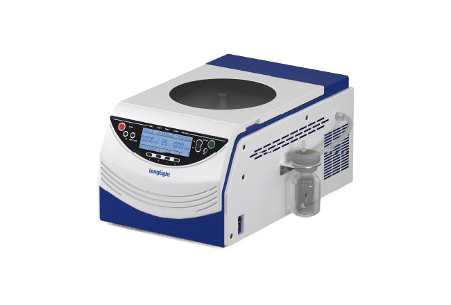
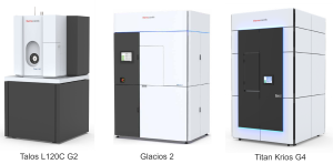
Three Major Equipment
Our cryo-EM services are supported by access to high-end scientific research platforms equipped with state-of-the-art instrumentation.
Available systems include Talos L120C G2, an entry-level transmission electron microscopy and cryo-EM screening platform; Glacios 2, a 200 kV workhorse cryo-EM system optimized for routine single-particle and tomography workflows; and Titan Krios G4, a flagship 300 kV cryo-EM platform designed for maximum stability, automation, throughput, and resolution potential.
These platforms collectively support cryo-grid screening, single-particle analysis, cryo-electron tomography, and advanced method development across a wide range of structural biology applications.
Why Longlight?
We offer valuable and mature experience in cryo-electron microscopy through access to high-end scientific research platforms and collaboration with industry experts and academic scientific advisors. Our services emphasize full transparency and completeness of data delivery, covering raw data, intermediate processing files, final density maps, atomic models, and validation reports.
By combining advanced instrumentation, experienced data processing workflows, and balanced delivery timelines, we provide reliable, high-quality cryo-EM structural solutions tailored to diverse research needs.
Frequently Asked Questions.
Q1: How much sample do customers need to prepare?
- Negative stain:
minimal concentration ~1 g/L
minimal volume ~100 µL.
- SPA with soluble proteins:
minimal concentration ~1 g/L
minimal volume ~100µL.
- SPA with membrane proteins:
minimal concentration ~1 g/L
minimal volume ~100µL.
to be discussed and adjusted if needed
Q2: What are the requirements for sample buffer?
- pH range: 6.0-8.5
- salt concentration <200 mM
- low glycerol (how much? doi: 10.1107/S2053230X24002553)
- low azide (how much? supporting reference is missing)
Q3: What is a typical service process?
Project consultation → NDA signing → service agreement confirmation → sample receiving → quality inspection → negative stain → data collection → data processing → report delivery.
Q4: What is the average delivery time?
- Negative staining: 1-2 weeks.
- SPA, preliminary results: 6-8 weeks .
- SPA, high-resolution model: 2-3 months.
ChIP-seq
ChIP studies the interaction between DNA and proteins, and ChIP-seq is combined with next-generation sequencing to detect DNA sites in the genome that bind to specific transcription factors/histones, which is used to study the interaction between proteins and chromatin.
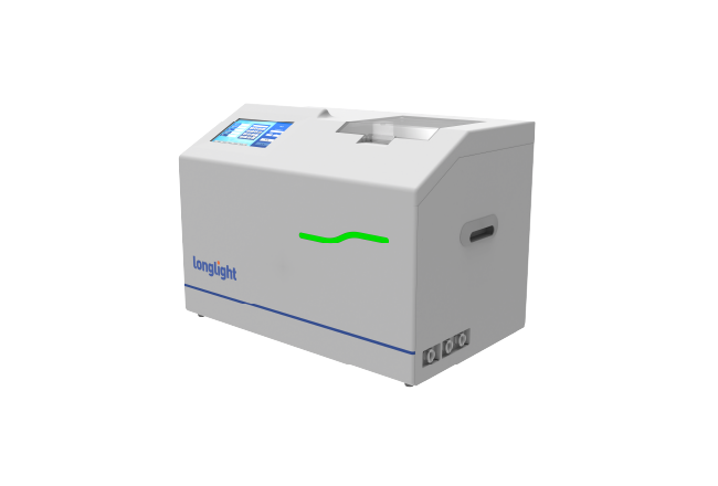
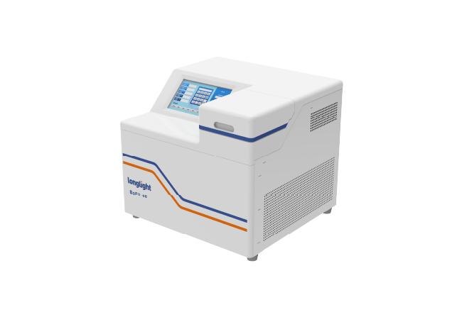
Genomics
Longlight has focused on molecular diagnosis and molecular biology, we has launched some NGS related instruments and reagent consumables, mainly Focused Ultrasonicator, been committed to promoting the development of genomics, to serve the human medical cause better.
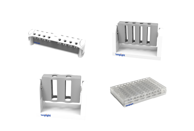
Consumables and Kits
Longlight provides different types of precast agarose gel, nucleic acid scavenger, Qubit tube, nucleic acid extraction kits, and library preparation kits. In the meanwhile, all of these products can be widely used in basic scientific research, biopharmaceuticals and other application scenarios.
Contact Us


Hotline Support
For faster response, you can contact us via phone:+86 0755-86727654