Related Post

Say Goodbye to Blurry Gel Electrophoresis Image Capture Issues
2025-11-27Gel Electrophoresis Image Capture records the bands formed after DNA, RNA, or proteins migrate through a gel. It turns invisible molecules into clear, auditable data. Labs use it for DNA profiling, CRISPR validation, and cancer biomarker studies. It supports microbial typing and vaccine research. Landmark projects, from the Human Genome Project to large COVID-19 studies, relied on precise gel images. Yet many teams still battle blur, noise, and weak bands. Why do good experiments produce bad pictures – and how can we fix them fast? Keep reading to see the simple steps that change everything.
(Band-collision gel electrophoresis | Nature Communications)
Why Blurry Images Happen – And Why It Matters
Blurry gels are not just an aesthetic issue. They lead to misreads, repeat runs, and shaky conclusions. In many labs, the root causes are familiar: low light output from dyes; cameras that drown weak bands in noise; shaky focus; and software that forces too much manual editing. Add inconsistent user handling and poor traceability, and it becomes hard to compare runs across time or across users.
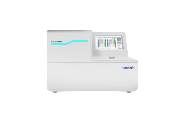
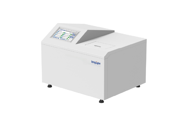
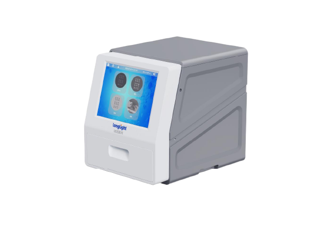
Longlight Technology addresses these pain points at the source. A high-sensitivity black-and-white CMOS camera captures fine band detail, even when signal levels are low. With 6.3-megapixel imaging and a 66 dB signal-to-noise ratio, faint bands stand out cleanly instead of merging into the background. Optical precision is matched by a stable illumination suite – trans-UV, trans-blue, and trans-white – so the system adapts to different dyes and stain-free workflows without compromise.
But clarity is not only about optics. Teams also need consistent handling. Role-based user management, categorized log viewing, and a full audit trail make it simple to see who did what, when, and how. That traceability reduces disagreement in group reviews and strengthens data integrity for audits and publications.
How Longlight Technology Delivers Sharp, Repeatable Results
Our philosophy is simple: make Gel Electrophoresis Image Capture fast, stable, and safe – without trading off accuracy.
- Smart Software, Less Rework
Our self-developed imaging software reduces tinkering and re-runs. Auto exposure and auto focus lock onto the optimal settings quickly. Image enhancement clarifies bands without distorting quantitation. Marker auto-annotation recognizes the ladder and labels band sizes automatically, which cuts down on hand edits and improves between-user consistency. The result is a cleaner analysis path from gel to report.
- Safe And Flexible For The Bench
A 12.1-inch touch display brings smooth, onboard processing right to the instrument, minimizing trips to a separate computer. The image area – 175 mm x 230 mm – comfortably covers common gel formats. External gel cutting is supported, and a UV-shield cutting plate blocks harmful UV during excision to protect users. The system runs on standard lab power (100 – 240 V, 50/60 Hz, 2.5 A) and fits easily on a bench with a compact footprint (about 465 x 357 x 425 mm). Compatibility with mainstream upstream and downstream tools keeps your current protocols intact.
You can tailor illumination to the chemistry at hand. Use trans-UV for classic ethidium bromide workflows, trans-blue for safer dyes, or trans-white for documentation and colony work. Switching is fast, so you can move from nucleic acid gels to protein gels or stain-free gels without reconfiguring your setup.
✅ Key Enhancements For Gel Electrophoresis Image Capture
- High-sensitivity 6.3-MP CMOS with 66 dB SNR for clear, intact bands under low light
- Trans-UV, trans-blue, and trans-white sources for broad dye compatibility
- Auto exposure, auto focus, and image enhancement to reduce manual tuning
- Marker auto-annotation to label ladder sizes and cut editing time
- Role-based users, logs, and full audit trail for compliance and reproducibility
- UV-shield cutting plate and external gel cutting support for safer excision
- 12.1-inch touch display for all-in-one operation and faster decisions
What You Gain?
When Gel Electrophoresis Image Capture becomes reliable, everything downstream improves. Teams finish runs on schedule, skip avoidable repeats, and share results with confidence. Quantitation is steadier because weak bands are still readable, and file naming, user tracking, and logs keep reviews efficient. For busy labs, these small gains add up to shorter cycle times and fewer weekend reruns.
Applications span nucleic acid imaging, SDS-PAGE protein gels, multi-color fluorescent labeling, stain-free workflows, and colony imaging. Switching between modes is straightforward, so the same instrument supports teaching labs in the morning and advanced fluorescence work in the afternoon. That versatility lowers total cost while raising utilization.
✅ Everyday Wins You’ll Notice
- Clear bands without long exposure trials, even for low-abundance targets
- Fewer manual annotations and re-captures; more time for analysis
- Consistent results across users, thanks to standardized software and audit trails
- Safer gel excision with effective UV shielding and external cutting support
At Longlight Technology, we believe instrumentation should disappear into your routine while lifting your results. If you are ready to say goodbye to blurry Gel Electrophoresis Image Capture, let’s talk. CTA: Request a live demo or a sample-data evaluation, and see how clear imaging, smart automation, and safer handling can raise your lab’s confidence from the first band to the final report.