Related Post
High Resolution Gel Imaging Explain: UV-Safe Gel Excision
2026-01-19High Resolution Gel Imaging now underpins many landmark advances in molecular biology. Teams led by Jennifer Doudna and Emmanuelle Charpentier relied on precise gel electrophoresis and high-resolution imaging to validate CRISPR constructs. Svante Pääbo's pioneering ancient DNA work demanded sensitive band detection to differentiate authentic fragments from contamination. The Human Genome Project, driven by groups including Eric Lander's team, scaled gel analysis to map and verify sequences. Across these efforts, one principle stayed constant: clean, well-resolved bands and gentle handling are essential. Today, UV-safe gel excision pairs with High Resolution Gel Imaging to protect both samples and staff while preserving downstream performance.
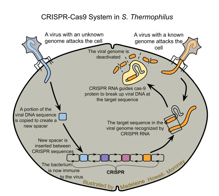
(Jennifer Doudna and Emmanuelle Charpentier's Experiment
About the CRISPR/cas 9 System's Role in Adaptive Bacterial Immunity (2012))
What Is High Resolution Gel Imaging
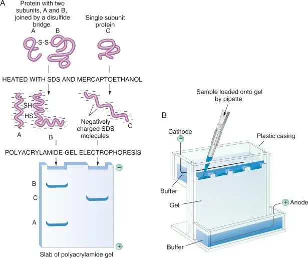
High Resolution Gel Imaging describes a set of optical, electronic, and software capabilities that capture crisp, high-contrast bands from nucleic acid or protein gels. It combines a sensitive camera, low-noise optics, matched filters, and calibrated illumination with intelligent image processing. The result is clear discrimination between close bands, accurate sizing, and reproducible quantitation, even when signals are weak or backgrounds are uneven. In practice, that yields reliable cloning, cleaner library prep, fewer repeats, and records suitable for regulated audits.
Beyond image clarity, the workflow is integrated end to end. Role-based access, audit trails, and automatic marker detection cut human error and bolster traceability. Support for multiple light sources and dyes expands applications from DNA and RNA gels to SDS-PAGE, stain-free protein workflows, colony imaging, and multi-color fluorescent labeling. High Resolution Gel Imaging has evolved from a camera-on-a-box to a connected, compliant instrument at the center of the bench.
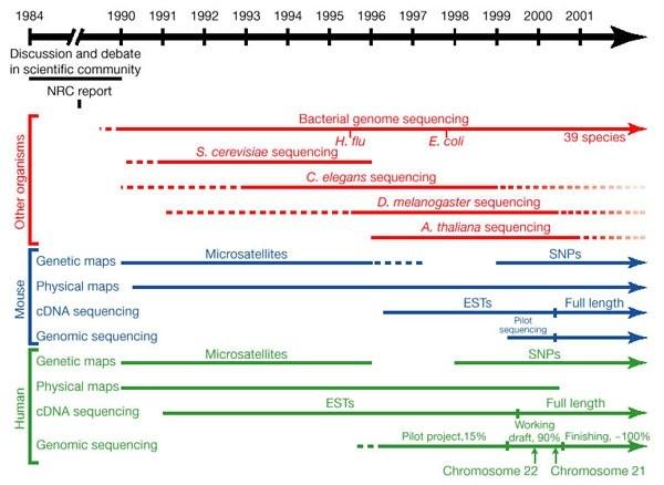
(Initial sequencing and analysis of the human genome | Nature)
Why UV-Safe Gel Excision Matters
Traditional UV transillumination can damage nucleic acids through thymine dimer formation and nicking. The consequences appear later: lower cloning efficiency, uneven library yields, and ambiguous Sanger or NGS results. UV exposure also poses a safety risk for users, complicating compliance and training.
Blue-light-based gel excision offers a safer path. By exciting common dyes without the high-energy wavelengths that harm DNA, blue light preserves fragment integrity during band cutting. When paired with a protective cutting surface that blocks UV, operators gain a clear view, steady hands, and confidence that both samples and people are protected. For labs under pressure to improve first-pass success, UV-safe excision is a direct lever to improve outcomes.
Typical pain points this approach solves:
• Bands smear or vanish after gel excision due to UV exposure
• Cloning success fluctuates even from the same lane
• Re-runs triggered by ladder labeling errors waste time
• Safety concerns and limited availability of UV stations
• Gaps in records complicate audits and quality inquiries
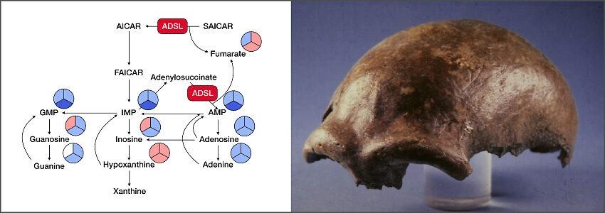
(Human Evolutionary Genomics Unit (Svante Pääbo) | OIST Groups)
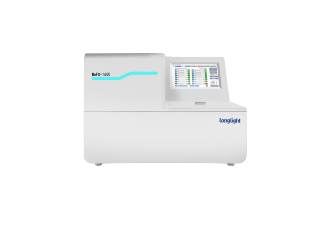
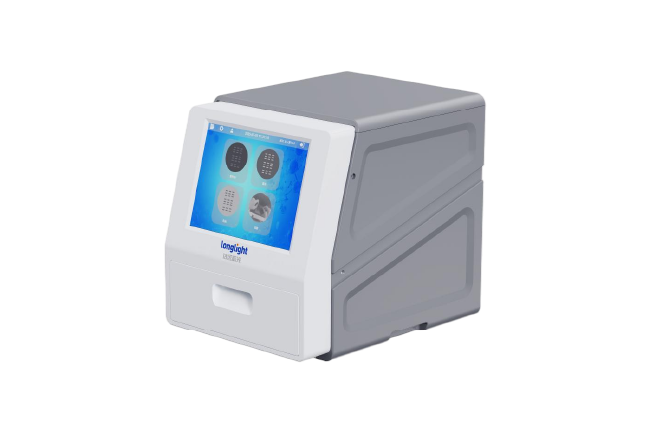
Inside Longlight High Resolution Gel Imaging System
Today's platform brings optics, control, analysis, and safety together in a single unit. Key elements include:
• All-in-one operation: With a 12.1-inch touchscreen and onboard processing, acquisition, tuning, and export happen at the bench - no external computers or cables required.
• Sensitive capture: A 6.3-MP high-sensitivity CMOS with low-noise optics reveals weak bands from limited-abundance samples on a uniform background. Fine distinctions are easier to see, supporting better selection and quantitation.
• Intelligent image processing: Automated exposure, autofocus, and enhancement deliver reproducible results. Marker auto-annotation detects ladder band sizes and applies labels, cutting down manual edits and avoiding transcription mistakes.
• Versatile illumination: Transmissive UV, blue, and white sources accommodate a wide range of dyes. One instrument unifies nucleic acid gel imaging, SDS-PAGE, stain-free protein detection, colony imaging, and multi-color fluorescence workflows.
• Data integrity and compliance: Role-based access, orderly log viewing, and a comprehensive audit trail secure full traceability across the workflow, reducing risk and making reviews easier for compliant or collaborative groups.
• UV-safe excision support: External gel excision is performed using a standard UV-shield cutting plate that blocks UV effectively, protecting users and supporting precise band isolation under blue light.
High Resolution Gel Imaging is not only about pixels. It is about a repeatable, documented workflow that turns a gel into defensible data and intact, ready-to-use DNA or protein samples.
From Imaging to UV-Safe Gel Excision: a Practical Workflow
Adopt the following sequence to align image quality with sample integrity:
• Prepare the gel and select a dye compatible with blue-light excitation to support UV-safe cutting.
• Place the gel on the stage and select the appropriate illumination. Start with blue light when excision is expected.
• Use automatic exposure and autofocus to capture a sharp, high-contrast image. Confirm that faint bands are visible without overexposure.
• Enable marker auto-annotation to label ladder band sizes and lock sizing parameters for downstream reporting.
• Switch to the external cutting surface with the UV-shield in place. Blue-light illumination preserves nucleic acids while providing clear visibility.
• Excise the chosen band with a clean scalpel; work fast and avoid re-lighting the gel.
• Document the removal with a rapid post-cut photo to secure traceability.
• Export images, metadata, and the comprehensive audit trail to LIMS or shared storage to wrap up.
This concise SOP reduces rework, shortens ramp-up, and produces consistent, audit-compliant records. It also unifies band selection across users, raising reproducibility across programs and timeframes.
Partner for Precision and Safety
Research teams today seek three outcomes from gel imaging: crisp, interpretable bands; intact samples after cutting; and reliable, searchable records. Our High Resolution Gel Imaging platform addresses each outcome in one system. It combines a high-definition optical engine with intelligent software, tri-mode illumination, and UV-safe gel excision support. It integrates smoothly with mainstream laboratory products and workflows, so you can apply it to nucleic acid and protein studies without disruption.
If your lab faces inconsistent band quality, low cloning yield after gel extraction, or audit gaps from manual annotations, consider a safer, smarter path. Discover High Resolution Gel Imaging and UV-Safe Gel Excision: protect samples and staff, switch to blue-light cutting, and get crisp bands with safer workflows. Contact us to request a demo, receive a sample image set from your dye of choice, or map the workflow to your SOPs with our applications team. By unifying image clarity, sample protection, and data integrity, you can move from troubleshooting to throughput - and keep your science, and your people, safe.