Related Post
RNA Electrophoresis Workflow Acceleration Guide| One-Step from Capture to Image Output
2026-01-16As a proven technique behind landmark discoveries, RNA electrophoresis is still the baseline for RNA sizing, integrity assessment, and processing profiling. From Andrew Z. Fire and Craig C. Mello's 1998 demonstration of RNA interference, where RNA electrophoresis and Northern blotting helped track double-stranded and small interfering RNAs, to Victor Ambros, Rosalind Lee, and Rhonda Feinbaum's 1993 identification of the lin-4 microRNA using polyacrylamide RNA electrophoresis, this technique has anchored modern molecular biology. Earlier, Phillip A. Sharp and Richard J. Roberts' work on RNA splicing, and Thomas R. Cech's studies on self-splicing introns, used gel-based analysis to resolve spliced products and RNA intermediates.
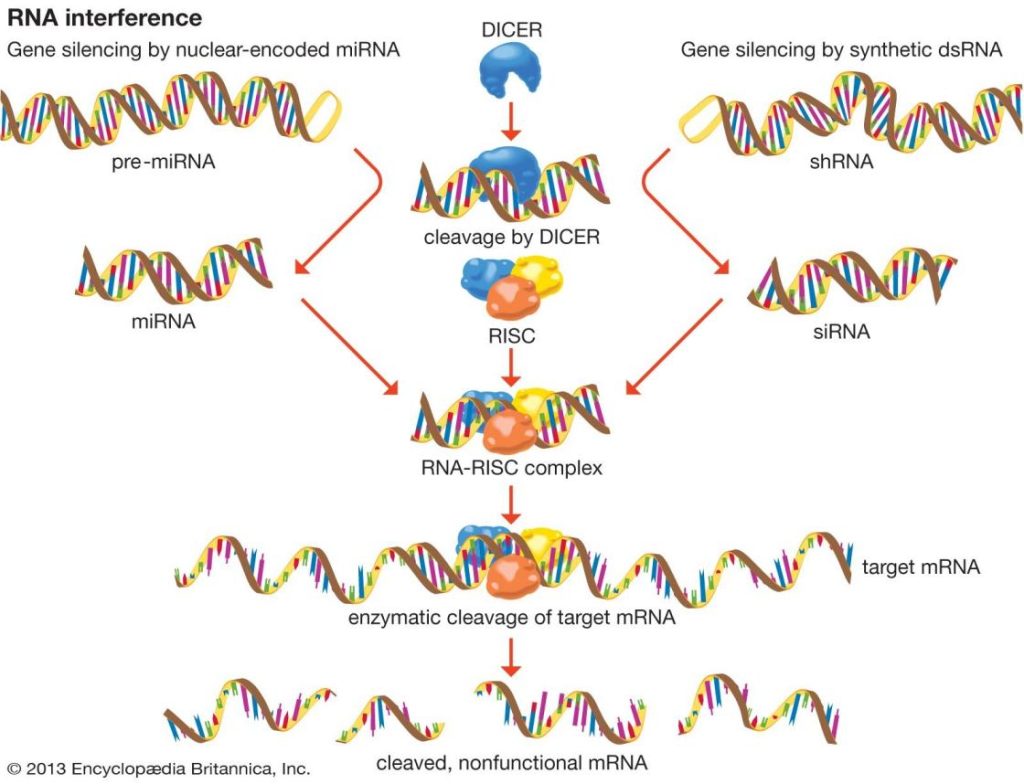
(Andrew Z. Fire | Nobel Prize-Winning American Geneticist | Britannica)
At Longlight Technology, we build systems that accelerate this essential workflow so researchers can move from gel to decision faster, with consistent quality and traceable data.
What Is RNA Electrophoresis?
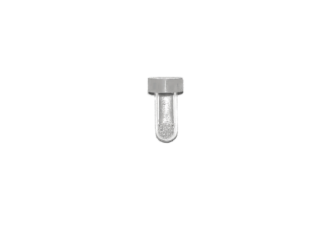
RNA Electrophoresis separates RNA by size through agarose or polyacrylamide gel matrices driven by an electric field. It enables viewing ribosomal RNA bands for integrity checks, flags degradation, and resolves small RNAs. The method also feeds Northern blotting by preparing size-fractionated RNA for hybridization. In routine laboratories, RNA electrophoresis validates sample quality prior to RT-qPCR, RNA-seq, or library construction, and confirms workflow steps such as fragmentation or adapter ligation.
Modern practice requires clear images in dim settings, wide-ranging dye/stain compatibility, and trustworthy quantitation. Given the sensitivity of RNA to RNases and UV light, careful regulation of imaging conditions is indispensable. Longlight Technology delivers a consolidated gel imaging platform tailored to RNA Electrophoresis and broader nucleic acid use, integrating accurate optics, automated processing, and safety-first handling in a compact footprint.
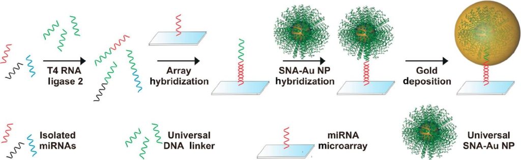
(MicroRNA: Function, Detection, and Bioanalysis | Chemical Reviews)
Industry Pain Points in RNA Electrophoresis
Many teams encounter shared bottlenecks when imaging RNA gels:
• Fragmented workflow. Different modules for excitation, optics, and software force handoffs and delays.
• Manual ladder annotation. Band-size labeling by hand takes time and can be subjective.
• Low-light sensitivity. Weak bands from low-abundance RNAs are obscured or missed amid noise.
• Safety risks. UV exposure during gel excision increases user hazard and demands rigorous shielding.
• Data traceability. Audit trails are incomplete, user roles are unclear, and logs are scattered.
• Compatibility gaps. Dye, light source, and software mismatches interrupt standard protocols.
The result is slower experiments, uneven documentation, and potential compliance risk. The solution is an integrated, one-step path that preserves quality while removing repetitive tasks.
Longlight Technology's One-Step Workflow - from Capture to Image Output
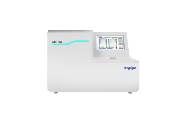
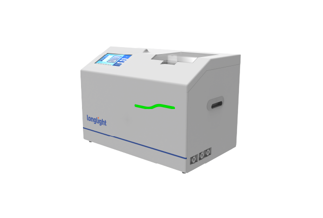
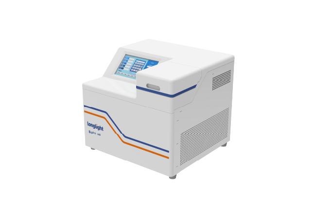
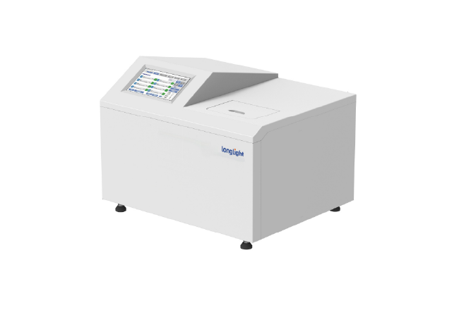
Longlight Technology designed an all-in-one Gel Imaging System to streamline RNA electrophoresis from capture to image output. The objective is simple: reduce clicks, increase clarity, and maintain traceability in a single pass.
- Capture
A high-sensitivity optical system pairs with transmissive UV, blue, and white light sources to support a broad range of RNA dyes. The 6.3-MP high-sensitivity CMOS camera records clear, intact bands under low-light conditions, improving detection of low-abundance RNAs. Automatic exposure and automatic focus stabilize imaging without manual tuning.
- Process
Onboard image processing runs directly on the instrument. A 12.1-inch touch display supports smooth control, image enhancement, and rapid review without external computers. This minimizes file transfers and reduces processing variability across operators. The interface is built for repeatable SOPs.
- Annotate and Trace
Marker auto-annotation recognizes ladder band sizes and labels them automatically, cutting manual edits and operator bias. Defined user roles, filterable log categories, and an end-to-end audit trail deliver traceability for every dataset across teams. Results are paired with consistent metadata, streamlining internal oversight and compliance obligations.
With this one-step workflow, RNA electrophoresis proceeds from gel placement to final annotated image on a single device. No toggling between platforms, no manual ladder drawing, and no lost logs.
Technical Highlights for RNA Electrophoresis Imaging
Longlight Technology builds systems to meet stringent lab demands while lowering total cost of ownership. Key capabilities for RNA electrophoresis include:
• High sensitivity optics. The 6.3-MP CMOS camera reduces background noise and captures weak signal bands, ideal for small RNAs and low-input samples.
• Intelligent image processing. Self-developed software supports automatic exposure, automatic focus, and image enhancement at the point of capture. Click once to acquire optimized results.
• Versatile applications. Support for UV, blue, and white transmissive light, plus fluorescence workflows, ensures broad dye compatibility across nucleic acid gels.
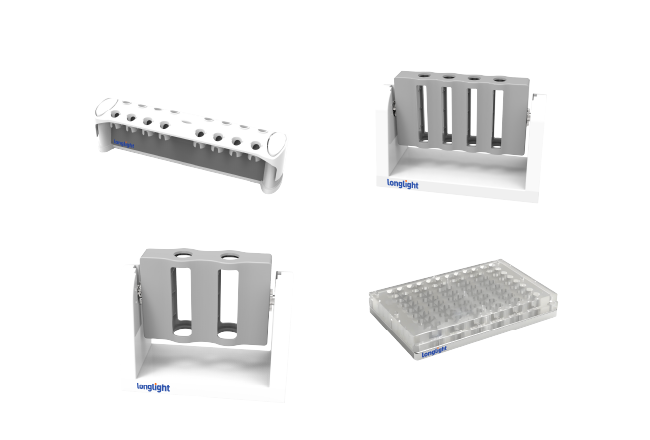
• Safety for gel excision. External gel cutting is supported with a standard UV-shield cutting plate that effectively blocks UV to protect users during band excision.
• All-in-one design. A 12.1" touch interface centralizes run control, imaging, and analysis for a streamlined user experience.
• Data management and traceability. Role-based governance, complete audit trails, and categorized logging uphold integrity and simplify regulatory reporting.
• Marker auto-annotation. Automatic marker annotation reduces manual steps and strengthens reproducibility.
• Compatibility. Ready integration with leading reagents, stains, and analysis tools supports nucleic acid and protein research across Longlight Gel Monitor systems.
These capabilities streamline sensitivity, speed, safety, and compliance, cutting onboarding time and standardizing outputs across teams working across sites.
Results, Compliance, and a Clear Call to Action
Implementing an integrated RNA electrophoresis approach produces faster turnaround, more consistent documentation, and safer bench workflows. Integrated capture-to-log on a single instrument prevents workflow fragmentation and data loss. In QC-rigorous operations, an end-to-end audit trail anchors validation and supports compliance.
• Single-step capture-to-report accelerates throughput.
• Clear, low-light imaging of small RNAs and compromised samples.
• Automatic ladder identification stabilizes size estimation and reduces human error.
• Role-based governance and complete audit trails enable study-level traceability.
• UV-shielded gel workstation improves safety for band excisions.
Longlight Technology's approach is built for modern molecular workflows. It helps RNA electrophoresis move at the pace of your research, not the pace of your instruments. If you are consolidating imaging platforms, scaling RNA QC, or seeking traceable documentation without adding complexity, we invite you to evaluate our one-step solution.
Call to Action
Contact Longlight Technology to request a demo and see how our all-in-one Gel Imaging System accelerates RNA electrophoresis - from capture to image output, with 12.1" touch control, onboard processing, auto labeling, and traceable data. Upgrade your gel imaging to a workflow that is faster, safer, and visibly more precise.